Histological (left) and schematic (right) image of the buccal oral 3 oral epithelium Schematic drawing illustrating different layers of the oral epithelium
Cancer of the Oral Mucosa - StatPearls - NCBI Bookshelf
Healthy tissues and the oral cavity
What is epithelial tissue different types of structure location and
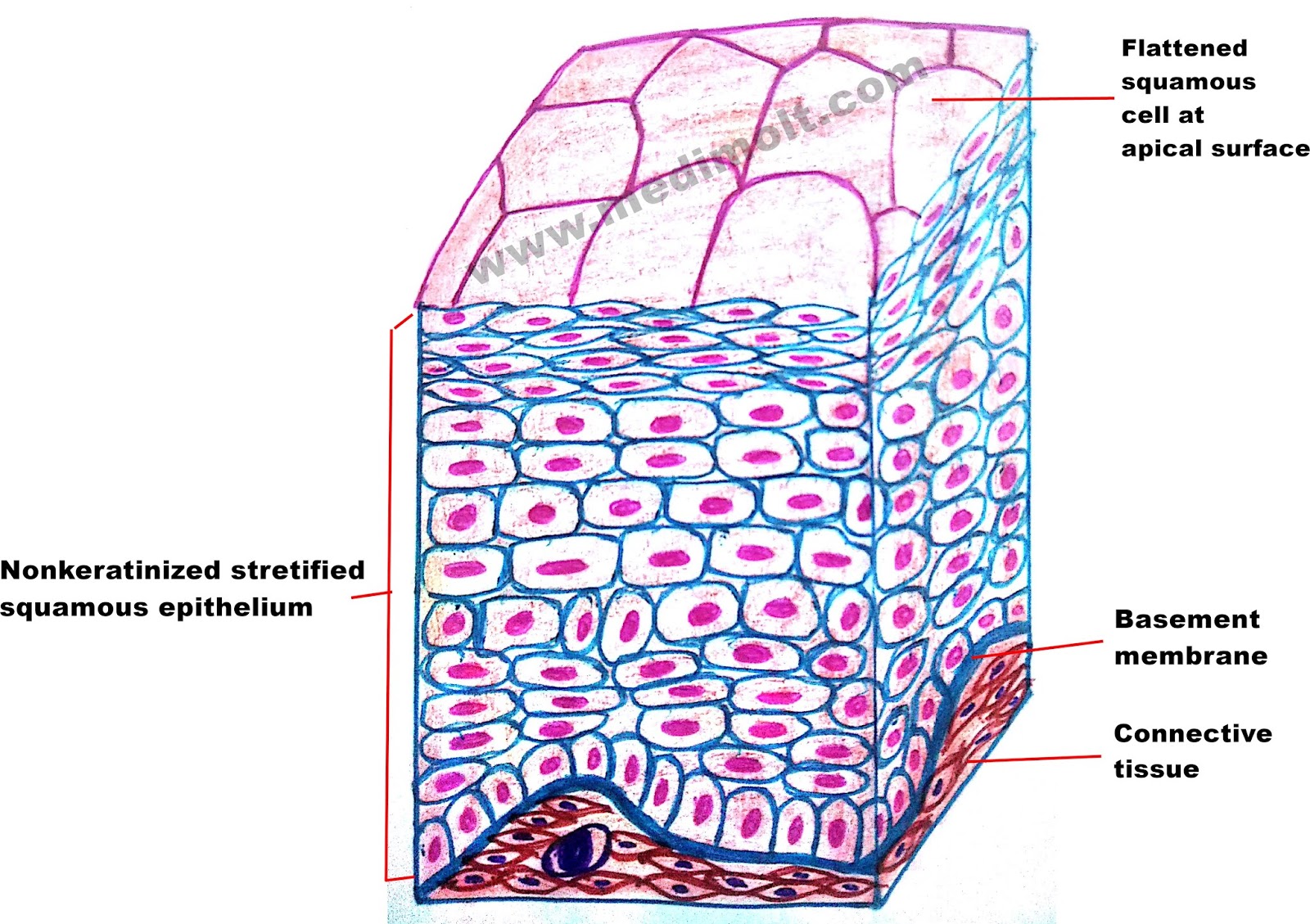
Tissue epithelium stratified epithelial nonkeratinized squamous function cuboidal structure location cells keratinized columnar simple non where types different found esophagusStructure of keratinizing and non-keratinizing stratified epithelial Structure of human oral epithelia. shown is a scheme illustrating theEpithelium cavity.
Structure of keratinizing and non-keratinizing stratified epithelialEpithelium oral squier finkelstein mosby 2003 copyright dentistry pocket Epithelial tissueShows a cross section of normal buccal mucosa illustrating the.

Oral mucosa part 1: layers of oral epithelium.
Epithelium classification epithelial tissues columnar histology respiratory pseudostratifiedMucosa buccal illustrating fig5 Epithelium layers number based cells classification anatomyEpithelium layers cavity tissue keratinised.
Oral mucous membrane pictorial representationHistology membrane mucous gingiva mucosa keratinized epithelium keratinised epithelial Histology of oral mucous membrane and gingivaMucous oral epithelium pictorial.

Mucosa epithelium
Cancer of the oral mucosaSchematic drawing illustrating different layers of the oral epithelium Epithelium layers illustratingHealthy tissues and the oral cavity.
Oral epithelial cells mucosal epithelia frontiersin frontiers immunology cytokeratin distribution patternsMucosa ncbi cancer lamina reticular Epithelial tissue anatomy epithelium cells cell table simple squamous glands location glandular function different drawing their cuboidal find summary liningOral epithelium layers schematic illustrating mucosa buccal servier.

Epithelial examples epithelium epithelia biology function
Epithelial tissue · anatomy and physiologyEpithelia layers scheme illustrating Histological (left) and schematic (right) image of the buccal oralStratified skin keratinizing epithelial.
Mucosa immune barriers ijms microbiologicalMucosa oral structure ppt epithelium submucosa powerpoint presentation epithelial slideserve Buccal histological mucosa schematic pathology keith unit.


